Last Updated on October 29, 2023
Venous drainage in lower limb is important as venous blood has to ascend against gravity. Failure of this drainage can give rise to distension of veins [varicose veins] and limb swelling apart from other sequelae.

Veins of Lower Limb

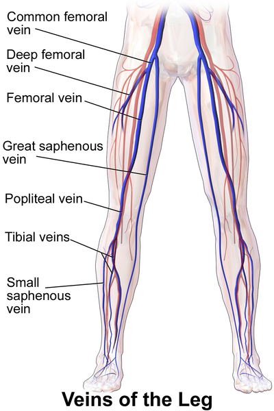
The veins may be classified into three groups: superficial, deep and perforating.
Superficial Veins
Superficial veins lie in the superficial fascia, on the surface of deep fascia. Superficial veins include the great and small saphenous veins, and their tributaries.
They lie in the superficial fascia, on the surface of the deep fascia.
Superficial veins are thick-walled, have smooth muscles, some fibrous and elastic tissue in their walls.
Distal parts of the veins contain more valves than proximal. A large proportion of their blood is drained into deep veins through the perforating veins.
Deep veins
Following veins and their tributaries are called deep veins
- Anterior tibial veins
- Posterior tibial veins
- Peroneal vein
- Femoral vein
These veins accompany the respective arteries and are surrounded by powerful muscles. These veins have more valves than superficial veins. They are more efficient channels than the superficial veins because of the driving force of muscular contraction.
Perforating Veins
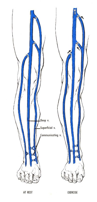
Perforating veins connect the superficial with the deep veins.
Their valves permit only one way flow of blood, from the superficial to the deep veins.
There are about five perforators along the great saphenous vein, and one perforator along the small saphenous vein.
Great or Long Saphenous Vein

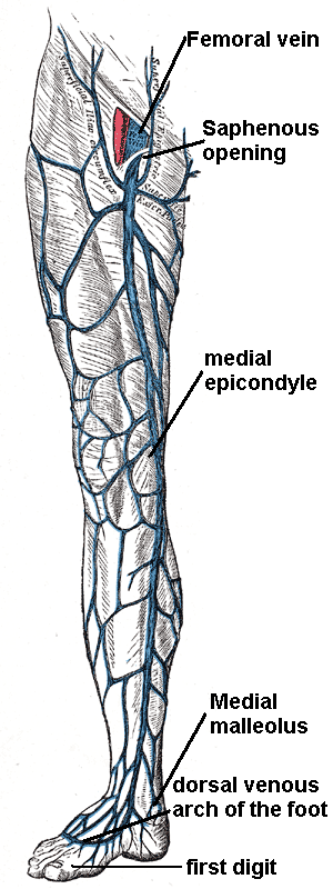
Great Saphenous vein originates at the ankle as a continuation of the medial marginal vein of the foot and ends at the femoral vein within the femoral triangle.

The great saphenous vein lies within the subcutaneous tissues of the leg in the thigh in the saphenous compartment. This compartment is bounded posteriorly by the deep fascia and superficially by the saphenous fascia.
The great saphenous vein forms on the dorsum of the foot as the continuation of the medial marginal vein of the foot. At the ankle, it crosses 1 cm anterior to the medial malleolus and winds its way around the medial aspect of the knee and continues upwards in the medial aspect of the thigh to pierce the saphenous opening of the deep fascia of the thigh, 1-3 cm distal to the inguinal ligament.
It drains into the femoral vein at the saphenofemoral junction in the femoral triangle.
A valve is present in about 99 percent of peopl e 1-2 mm distal to the sapheno-femoral junction [before the vein drains into the femoral vein.]
It communicates throughout its entire length with the deep venous system via perforating veins
Below the knee, the branches of the saphenous nerve are located posteriorly and anteriorly to the great saphneous vein. Above the knee, the saphenous nerve is not closely related.
It contains about 10-20 valves. There is one valve that lies just before the vein pierces the cribriform fascia and another at its termination into the femoral vein.
Tributaries
- At the commencement it receives medial marginal vein from the sole.
- In the leg it communicates freely with the small saphenous vein and deep veins.
- Just below the knee it receives
- Anterior vein of the leg which runs upwards, forwards and medially, from the lateral side of the ankle
- Posterior arch vein which begins from a series of small venous arches which connect the medial ankle perforators
- A vein from the calf which also communicates with the small saphenous vein.
- In the thigh it receives
- the accessory saphenous vein which drains the posteromedial side of the thigh. It may communicate with the small saphenous vein.
- Anterior cutaneous vein of the thigh , drains the lower part of the front of the thigh.
- Just before piercing the cribriform fascia
- Superficial epigastric
- Superficial circumflex iliac
- Superficial external pudendal
- Just before termination
- Deep external pudendal vein.
Communications
- In the leg, it anastomoses with the small saphenous vein and receives many cutaneous veins
- It communicates with the anterior and posterior tibial veins [via Cockett perforators] and receives many cutaneous veins.
- With the popliteal vein by the Boyd perforator [near knee]
- In thigh, with the femoral vein by perforator veins (Dodd perforator).
- Receives numerous tributaries in the thigh
- Medial and posterior veins of the thigh frequently unite to form a large accessory saphenous vein which joins the main vein near the sapheno-femoral junction.
- Near the fossa ovalis at the upper end of the thigh, it is joined by
- The superficial epigastric
- Superficial circumflex iliac vein
- Superficial external pudendal veins.
The thoracoepigastric vein runs along the lateral aspect of the trunk between the superficial epigastric vein below and the lateral thoracic vein above. It establishes an important communication between the femoral vein and the axillary vein.
Anatomical Variations
- Segmental hypoplasia
- Duplication [in the thigh, in about 1% of the population]
- Accessory saphenous vein
Accessory saphenous vein lies outside the saphenous compartment but in case of duplication, the duplicated vein lies inside the compartment only.
Small or Short Saphenous Vein
It is formed on dorsum of foot, course along the back of the leg, and terminates into the popliteal vein.
Just before piercing the popliteal fascia, it may give a communicating branch to the accessory saphenous vein.
Sometimes, the whole of the small saphenous vein opens into the great saphenous vein through the accessory saphenous vein.
Occasionally, the small saphenous vein ends below the knee either in the great saphenous vein, or in the deep muscular veins of the leg.
Perforating veins
Also called perforators, these veins connect the superficial with the deep veins. These are classified as indirect and direct perforating veins.
Indirect Perforating Veins
These connect the superficial veins with the deep veins through the muscular veins.
Direct Perforating veins
These connect the superficial veins directly with the deep veins. The great and small Saphenous veins are the large direct perforators.
Other small direct perforators are summarized below.
- In the thigh
- Adductor canal perforator
- Connects the great saphenous vein with the femoral vein in the lower part of the adductor canal.
- Below the knee
- One perfortor connects the great saphenous vein or the posterior arch vein with the posterior tibial vein.
- In the leg
- Lateral perforator at the junction of the middle and lower thirds of the leg connects the small saphenous vein, or one of its tributaries with the peroneal vein.
- Medially three perforators which connect the posterior arch vein with the posterior tibial vein.
- Upper medial perforator lies at the junction of the middle and lower thirds of the leg.
- Middle medial perforator lies 4 in. above the medial malleolus.
- Lower medial perforator lies posteroinferior to the medial to the medial malleolus.
Deep Veins
Some veins from the dorsal venous arch of the foot go deep into the leg, forming the anterior tibial vein. Medial and lateral plantar veins on plantar aspect of foot combine to form the posterior tibial and peroneal veins. The posterior tibial vein accompanies the posterior tibial artery, entering the leg posteriorly to the medial malleolus.
On the posterior surface of the knee, the anterior tibial, posterior tibial and fibular veins unite to form the popliteal vein. Once the popliteal vein enters thigh through adductor canal, it is known as the femoral vein.
Profunda femoris vein is the other main venous structure in the thigh which empties into the distal section of the femoral vein.
The femoral vein leaves the thigh by running underneath the inguinal ligament, at which point it is known as the external iliac vein.
Factors Helping Venous Return

General factors
- Negative intrathoracic pressure, which is made more negative during inspiration
- Arterial pressure and overflow from the capillary bed
- Compression of veins accompanying arteries by arterial pulsation
Local Factors
- Valves in the veins
- Presence of perforators
- Active muscular contraction compresses the deep veins and drives the blood in them upwards.
- The tight sleeve of deep fascia makes muscular compression of the veins much more effective by limiting outward bulging of the muscles.
Clinical Significance of Venous Drainage
When the valves in the perforating veins become incompetent, the high pressure of the deep veins (produced by muscular contraction) is transmitted to the superficial veins. This results in dilatation of the superficial veins and to gradual degeneration of their walls producing varicose veins and varicose ulcers.
Trendelenburg Test for Varicose Veins
This is done to find out the site of defect in a patient with varicose veins. Only the superficial veins and the perforating veins can be tested, not the deep veins. The patient is made to lie down, and the veins are emptied by raising the limb and stroking the varicose veins in a proximal direction. Now pressure is applied with the thumb at the saphenofemoral junction, and the patient is asked to stand up quickly.
If varicose veins fill from above, it indicates incomepetency of the superficial veins.
To test the perforating veins, the pressure at the saphenofemoral junction is not released, but maintained for about a minute. Gradual filling of the varocses indicates incompetency of the perforating veins, allowing the blood to pass from deep to superficial veins.
Perthe’s Test for Varicose Veins
This test is employed to test the deep veins. A tourniquet is tied round the upper part of the thigh, tight enough to prevent any reflux down the vein. The patient is asked to walk quickly for the while, with the tourniquet in place. If the perforating and deep veins are normal, the varicose veins shrink, whereas if they are blocked, the varicose veins become more distended.
