Last Updated on March 18, 2020
A tumor is a mass of abnormal tissue.
The female breast is composed of different types of tissues including glandular tissue, fatty tissue, and fibrous stroma. Breast tumors can arise from any of these tissues.
Read more about Anatomy of the Breast
Classification of Breast Tumors
Breast tumor can be of two types:
- Benign (non-cancerous) Tumors
- Malignant (cancerous) Tumors
It’s important to remember that most of the breast lumps are benign (meaning they are not cancerous) and not of serious consequence. But since a breast lump can also be due to cancer, one must seek medical help for any lump or swelling discovered in the breasts.

Difference Between Tumor, Neoplasm, Mass, and Cancer
In a purely technical sense, tumor refers to a neoplasm which means an abnormal new growth of cells which could be benign or malignant. It is actually a synonym for neoplasm.
But in routine usage, tumor refers to any mass or lump present within the breast. The mass or lump could be due to infection (e.g., breast abscess), cystic (e.g., simple breast cyst), benign tumor (e.g., fibroadenoma) or malignant tumor (e.g., breast cancer).
Read more about Breast Cancer: Risk Factors, Classification, Diagnosis and Treatment
In either case, the term tumor is not the same as cancer. Cancer means a malignant lesion or malignant tumor
In this article, we shall discuss the various types of breast tumors including benign and malignant ones. For other breast lumps or masses refer to the following article
Breast Lumps: Causes, Diagnosis, and Treatment

Image Credit: https://www.siyavula.com
Benign Breast tumors
Benign breast tumors are localized abnormal growths, but they do not invade the surrounding structures or spread beyond the breast. They are not life-threatening. They can be removed with surgery and do not usually come back (or recur).
The various benign tumors of the breast are:
Fibroadenomas
Fibroadenoma is one of the most common breast tumors in young women. It is a solid tumor composed of fibrous and glandular tissue.
It occurs most commonly in women between 15-35 years of age.
They are thought to occur due to the changing levels of hormones since they often appear during puberty or pregnancy and usually disappear after menopause.
One or several fibroadenomas can occur in a woman. They can be present in one or both breasts. Most fibroadenomas are small in size measuring about 1–2 cm, but they can grow as large as 5 cm or even more.
It presents as a lump in the breast. The lump feels rubbery, smooth with well-defined edges. It is usually painless and freely movable within the breast tissue on palpation (when rolled with pads of the fingers). If situated deep within the breast tissue, it may not be felt and is discovered only by investigations like ultrasound or mammogram.
It is a benign tumor and its presence does not increase the risk of developing cancer.
Treatment is by surgical removal (lumpectomy or removal of the entire lump). Very small lumps (less than 2-3 cm in size) however, may not be removed and simply observed. In many cases, it tends to resolve on its own. If it grows over time or results in a change in the shape of the breast, it should be removed.
Intraductal papilloma
These are wart-like tumors that grow within the milk ducts present in the breast. They are made up of glandular tissue along with blood vessels and fibrous tissue (called fibrovascular tissue).
They are usually situated close to the nipple but can be found elsewhere in the breast. It’s most common in women over 40 years of age.
They may be single (solitary intraductal papillomas) which often grow in the large milk ducts near the nipple. They may be felt as a small lump behind or near the nipple. They often produce clear or greenish or bloody discharge from the nipple. They may also cause pain or discomfort.
Multiple papillomas are present in small ducts away from the nipple. They do not usually cause nipple discharge.
Papillomatosis is a condition in which very small areas of cell growth are present within the ducts. These small growths, however, are not as distinct as papillomas.
Although intraductal papillomas are benign, multiple papillomas, have been associated with a slightly higher risk of breast cancer.
Treatment is by surgical excision in which the papilloma along with a part of the duct is removed.
Phyllodes tumor
Similar to fibroadenoma, phyllodes tumor is a biphasic tumor arising from the fibrous and glandular tissue of the breast. It is a rare tumor accounting for less than 1% of all breast tumors.
Most of the phyllodes tumors ( about 90%) are benign. Some are classified as malignant or borderline (meaning that their malignant potential is uncertain).
Phyllodes tumors tend to grow quickly, but they rarely spread outside the breast.
Phyllodes tumor produces a firm round lump. Rarely it can cause pain. All three kinds of phyllodes tumors including the benign ones often grow very quickly, and they may be of quite a large size when they are first brought to the doctor’s attention.
They can occur at any age but usually occur in a woman after 40 years of age. Benign phyllodes tumors usually occur at a younger age than malignant phyllodes tumors.
Treatment is by surgery which includes lumpectomy (removal of the lump) along with excising a wide area of normal breast tissue around the tumor (called wide surgical excision).
It is important to remove the adjacent breast tissue along with the lump otherwise these tumors can recur. Even malignant phyllodes are treated in a similar manner. Extremely large malignant phyllodes or malignant phyllodes which recur may be treated by mastectomy ( removal of the whole breast)
Other rare benign tumors of the breast
A variety of breast tumors can arise from the different tissues present in the breast including fatty tissue, blood vessels, nerves, fibrous tissue, etc. Definite diagnosis of all these is by biopsy and treatment is by surgical removal of the lump (lumpectomy). These include:
-
Lipoma
-
Neurofibroma
-
Schwannoma
-
Leiomyoma
-
Granular cell tumor
-
Hemangioma
-
Adenomas
-
Tubular adenoma
-
Lactating adenoma
-
Apocrine adenoma
-
Ductal adenoma
-
Malignant Breast Tumors (Cancer)
Malignant tumors of the breast, if not detected and treated early, continue to grow, invade and destroy the adjacent normal breast tissue. They have a tendency to spread to surrounding lymph nodes. If unchecked, the cancer cells tend to break away from the tumor and then through the lymphatic system and blood vessels spread to other areas of the body like liver, lung, bones, brain, etc. This process is called metastasis. Once cancer has metastasized, it is lethal, and chances of cure are very bleak as compared to the localized early stage. Hence it is important that all breast lumps be brought to medical attention as soon as possible and investigated properly. Once cancer, is diagnosed, it must be promptly and adequately treated.
Read more about Metastasis or Metastatic Disease
The 2 most common cancers arising from the glandular part of the breast are
-
Invasive ductal carcinoma
-
Invasive lobular carcinoma
Other less common forms include tubular carcinoma, cribriform carcinoma, papillary carcinoma, mucinous carcinoma, metaplastic carcinoma, etc.
Rare forms of cancer involving the breast include
-
Liposarcoma
-
Angiosarcoma
-
Rhabdomyosarcoma
-
Osteosarcoma
-
Leiomyosarcoma
-
Lymphoma
-
Malignant phyllodes tumor
Symptoms of breast cancer
- The most common symptom of breast cancer is a lump in the breast or the armpit which is usually hard and painless. The lump may be fixed to overlying skin which means that it does not move beneath the skin. It may even be attached to the underlying structures of the breast.
- There may be dimpling or redness or scaling and crusting of the skin of the breast or the skin may have a pitted appearance resembling an orange peel.
- Discharge from the nipple which could be bloody; inverted nipple and change in the shape or appearance of the nipple may also be the presenting signs of breast cancer.
Investigations of Breast Tumors

Image Credit: https://cancercentrelondon.co.uk
Mammogram
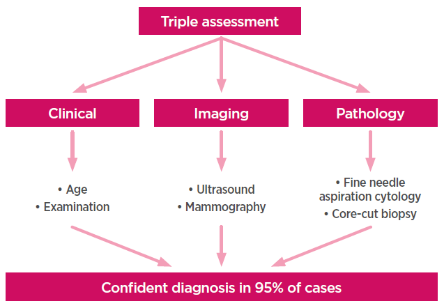
Mammography uses low dose x-rays to examine the breasts. The breasts are exposed to a small amount of ionizing radiation and pictures of the inside of the breasts from different angles are obtained. It is the most effective non-invasive test to detect breast cancer.
Read more about Screening of Breast Cancer
Breast ultrasound
Ultrasound uses high-frequency sound waves to obtain pictures of the inside of the breasts. It is non-invasive and free of harmful radiation. It can capture images of areas of the breast that are difficult to see with mammography. It can distinguish between fluid-filled (cystic) and solid breast lumps. During pregnancy and lactation, ultrasound is considered superior to mammography because the hormone-induced changes in breast tissue cause an increase in the density of breast tissue making interpretation of mammograms difficult.
Breast MRI (Magnetic Resonance Imaging)
MRI uses a powerful magnetic field, radiofrequency pulses and a computer to produce images of the inside of the breasts. A contrast material is injected through a vein into the body before obtaining the images. MRI can be used to diagnose breast lumps in select high-risk patients, in cases of dense breast tissue or when findings of mammogram and ultrasound are not conclusive.
Read more about MRI in Breast Cancer- Indications and Procedure
Nipple discharge cytology
The discharge from the nipple can be smeared on a glass slide and examined under a microscope to look for the presence of malignant cells.
Fine needle aspiration cytology (FNAC)
In this procedure, a fine needle is inserted into the lump and some material is aspirated. This material is then spread on a glass slide, stained with appropriate stains and examined under a microscope.
The procedure may be performed under the guidance of ultrasound.
In most of the cases, FNAC is able to categorize the breast lump as infectious, benign or cancerous. It is also a quick, relatively painless, non-invasive, outdoor procedure. The results are available within a few hours and can help to relieve the patient’s anxiety quickly.
Biopsy
Sometimes, when the results are inconclusive on FNAC, a biopsy may have to be performed to be sure that the breast lump is not cancerous. It is a small surgical procedure carried out under local anesthesia. A small piece of the breast lump is removed (and after a series of steps including processing, fixation, and staining) it is examined under a microscope. It is the best method to confirm whether cancer is present or not. However, the processing and other steps involved make it a lengthy procedure. The results are available in about 3-4 days.
A breast biopsy can be of various types
Core needle biopsy: A needle larger than that used for FNAC and having a special tip is used to remove a tiny sample of breast tissue.
Stereotactic biopsy: Mammography is used to precisely identify the suspicious area and then that area is biopsied.
Vacuum-assisted biopsy: A probe with a vacuum is inserted through a small incision in the skin and a sample of breast tissue is removed. It is performed under imaging guidance (ultrasound or mammogram or MRI).
Surgical biopsy: A small cut is made in the skin and part or whole of the lump is removed.
- Incisional biopsy: Only a part of the lump is removed.
- Excisional biopsy: The entire lump is removed. It is also called lumpectomy. If the lump turns out to be non-cancerous, it also serves as the only treatment required.
Treatment of Breast Tumors
Benign tumors
For the majority of benign breast tumors, surgical excision of the entire lump or lumpectomy results in a complete cure. Very small lumps (less than 2-3 cm in size ) however, may not be treated and kept under observation. Phyllodes tumors require surgical removal of the lump along with a margin of healthy tissue (wide surgical excision).
Malignant tumors (cancer)
Treatment depends on the stage of cancer. Staging is done on the basis of the size of the tumor, invasive or noninvasive nature of the tumor, lymph nodes involvement and spread of cancer to nearby or distant organs
The various treatment modalities include surgery (lumpectomy meaning removal of the lump or mastectomy meaning the removal of the breast), chemotherapy and radiotherapy.
