Last Updated on October 29, 2023
Anemia is defined as a decrease in the amount of red blood cells or hemoglobin in the blood which decreases the ability of the blood to carry oxygen.
Anemia isn’t a disease in itself, but a manifestation of malfunction somewhere in the body.
Anemia is the most common disorder of the blood affecting about a quarter of people all around the world. Iron-deficiency anemia affects nearly 1 billion. It is more common in females than males, among children, during pregnancy, and in the elderly.
Age-wise Hemoglobin Levels for Anemia
WHO’s hemoglobin thresholds used to define anemia |
||
|
Age or gender group |
Hb threshold (g/dl) |
|
| Children (0.5–5.0 yrs) | 11.0 | |
| Children (5–12 yrs) | 11.5 | |
| Teens (12–15 yrs) | 12.0 | |
| Women, non-pregnant (>15yrs) | 12.0 | |
| Women, pregnant | 11.0 | |
| Men (>15yrs) | 13.0 | |
Erythropoiesis – How Red Blood Cells are produced
Our body contains about 25 trillion red blood cells. About 0.00001% are destroyed daily. To just give you an idea, about 2.5 million red blood cells are being destroyed every second. In normal homeostasis, these cells are replenished by the production of new cells in the bone marrow.
RBCs have an average span of life of 120 days.
Production of new red blood cells is called erythropoiesis which is carried through the following stages.
Hemocytoblasts
These are stem cells in the bone marrow which would give rise to all blood cells.
Proerythroblasts
These are differentiated stem cells which would form red blood cells. These are formed by division and differentiation of hemocytoblasts.
Early Erythroblasts
During this stage in erythropoiesis, hemoglobin synthesis begins. These are also called as basophilic erythroblasts
Intermediate erythroblasts
Hemoglobin synthesis continues and hemoglobin starts accumulating
Late erythroblasts
The nucleus is extruded out from the cell.
Reticulocyte
Reticulocyte is a precursor of mature red blood cells. These are so named because they exhibit a net-like appearance or reticulum in their cytoplasm when stained. About 1-3% of reticulocytes are found in the circulation.
Mature erythrocytes
This stage is determined by loss of ribosomes. These cells enter the circulation.
Aging and Breakdown of Red Blood Cells
A typical mature erythrocyte lives for about 120 days in the circulation. Red blood cells lack nucleus and cannot divide or synthesize new cellular components. Therefore, these cells degenerate due to aging or damage.
Old damaged RBC’s are removed from the circulation in the following ways:
- Phagocytic activities of macrophages in the liver, spleen and lymph nodes remove about 90% of red blood cells
- 10% of the old cells hemolyze in the circulation and their fragments are engulfed by macrophages.
Control of Erythropoiesis
A reduction in the oxygen levels or oxygen-carrying capacity is the stimulator for erythropoiesis. This reduction in oxygen may occur due to bleeding, exposure to high altitude, hemolytic disease resulting in low hemoglobin levels.
Hypoxia in blood specifically stimulates the juxtaglomerular apparatus within the nephron of the kidney to produce and release erythropoietin.
Erythropoietin stimulates the red marrow of the bones to form increased numbers of proerythroblasts and hasten the maturation of red blood cells. The increased concentration of red blood cells leads to the increased oxygen-carrying capacity of the blood which in turn produces a negative feedback control on red blood cell production.
Causes of Anemia
Impaired Production of Red Blood Cells
- Disturbance of proliferation and maturation of erythroblasts
- Iron deficiency anemia
- Pernicious anemia due to vitamin B12 deficiency –megaloblastic anemia
- Anemia of folic acid deficiency also causes megaloblastic anemia
- Anemia of prematurity, in premature infants at two to six weeks of age.
- Thalassemias
- Congenital dyserythropoietic anemias
- Anemia of renal failure
- Disturbance of proliferation and differentiation of stem cells
- Pure red cell aplasia
- Aplastic anemia
- Anemia of renal failure
- Anemia of endocrine disorders
- Other mechanisms of impaired RBC production
- Myelophthisic anemia or myelophthisis resulting from the replacement of bone marrow by malignant tumors or granulomas.
- Myelodysplastic syndrome
- Anemia of chronic inflammation
Increased destruction of Red Blood Cells (Hemolytic Anemias)
- Intrinsic (intracorpuscular) abnormalities cause premature destruction
- Hereditary spherocytosis
- Hereditary elliptocytosis
- Abetalipoproteinemia,
- Enzyme deficiencies
- Hemoglobinopathies
- Sickle cell anemia
- Hemoglobinopathies causing unstable hemoglobins
- Paroxysmal nocturnal hemoglobinuria
- Extrinsic (extracorpuscular) abnormalities
- Antibody-mediated
- Warm autoimmune hemolytic anemia
- Cold agglutinin hemolytic anemia
- Rh disease
- Transfusion reaction to blood transfusions
- Mechanical trauma to red cells
- Microangiopathic hemolytic anemias
- Infections, including malaria
- Heart surgery
- Haemodialysis
- Antibody-mediated
Increased Blood Loss
- Trauma or surgery, causing acute blood loss
- Gastrointestinal tract lesions
- Intestinal parasitic infestation
- Gynecologic disturbances
- Excess menstrual blood loss
- Anemia of prematurity
Fluid overload (Hypervolemia)
- Excessive sodium or fluid intake, sodium or water retention
- Anemia of pregnancy
Classification of Anemia
Kinetic classification of anemia
The number of RBCs present in the circulation is the result of an equilibrium between production and delivery of red cells into the circulation and their destruction or loss from the circulation. Enhanced red cell production is indicated by an increase in circulating reticulocytes. (precursor of mature RBCs)
Reticulocyte production index < 2
- Iron deficiency
- Anemia of chronic disorders
- Aplastic anemia
- Renal disease
- Megaloblastic anemia
- Thalassemia
Reticulocyte production index > 2
- Hemolytic anemias
- Treated nutritional anemias
Morphological classification of anemia
Anemia is classified by the size of red blood cells. The size is reflected in the mean corpuscular volume (MCV). Microcytic anemia is often the result of iron deficiency. Limitations of MCV include cases where the underlying cause is due to a combination of factors – such as iron deficiency (a cause of microcytosis) and vitamin B12 deficiency (a cause of macrocytosis) where the net result can be normocytic cells.
 Microcytic Anemia
Microcytic anemia occurs as a result of failure or decreased hemoglobin synthesis.
- Heme synthesis defect
- Iron deficiency anemia
- Anemia of chronic disease
- Globin synthesis defect
- Alpha-, and beta-thalassemia
- HbE syndrome
- HbC syndrome
- Unstable hemoglobin diseases
- Sideroblastic defect
- Hereditary sideroblastic anemia
- Acquired sideroblastic anemia, including lead toxicity
Iron deficiency anemia is the most common type of anemia worldwide. Causes of iron deficiency include:
- Insufficient dietary intake or absorption of iron to meet the body’s needs. Infants, toddlers, and pregnant women have higher than average needs. Iron is an essential part of hemoglobin, and low iron levels result in decreased incorporation of hemoglobin into red blood cells.
- Blood loss from the gastrointestinal tract due to colon polyps or colorectal cancer. Fecal occult blood testing, upper and lower endoscopy should be performed to identify these.
- Intestinal parasitic infestation.
Read more about Iron Deficiency Anemia: Causes, Lab diagnosis, and Treatment
Macrocytic Anemia
By means of morphological and biochemical criteria, macrocytic anemia can be further divided into megaloblastic and nonmegaloblastic anemia.
- Megaloblastic anemia is due to a deficiency or reduced absorption of either vitamin B12, folic acid, or both. Pernicious anemia is caused by a lack of intrinsic factor, which is required to absorb vitamin B12 from food.
- Nonmegaloblastic anemia occurs due to hypothyroidism, alcoholism and liver diseases.
The cause of megaloblastic anemia is primarily a failure of DNA synthesis with preserved RNA synthesis, which results in restricted cell division of the progenitor cells. Specific features of vitamin B12 deficiency include peripheral neuropathy and subacute combined degeneration of the cord, a smooth, red tongue, and glossitis.
Read more about Megaloblastic anemia: Causes, Pathophysiology, Investigations, and Treatment
Read more about Pernicious Anemia: Causes, Investigations, and Treatment
Normocytic Anemia
Normocytic anemia occurs when MCV and MCHC are within normal limits. They include disorders having diverse pathogenetic mechanisms such as:
- Posthaemorrhagic anemia
- Anemia of chronic disease
- Aplastic anemia
- Hemolytic anemia
- Anemia of renal insufficiency
Dimorphic Anemia
A dimorphic appearance on a peripheral smear occurs when there are two populations of red blood cells, Causes include:
- Sideroblastic anemia
- A person recently transfused for iron deficiency or folate or vitamin B12 deficiency- Two populations of RBCs would be seen- donor RBCs of normal size and color and recipient RBCs which would be either small and pale (microcytic hypochromic) or larger (macrocytic) than normal.
Symptoms and Signs of Anemia
Mild anemias may get unnoticed. The symptoms of anemia may be related to underlying causes or the anemia itself. Common symptoms include weakness, fatigue, general malaise, poor concentration and shortness of breath.
Chronic anemia may result in behavioral disturbances in children and reduced scholastic performance in school-going children.
Pica, the consumption of non-food items such as dirt, ice, mud, paper, wax, grass, maybe a symptom of iron deficiency.
In very severe anemia, the body may compensate for the lack of oxygen-carrying capability of the blood by increasing cardiac output. The patient may then have symptoms such as palpitations, angina, and heart failure.
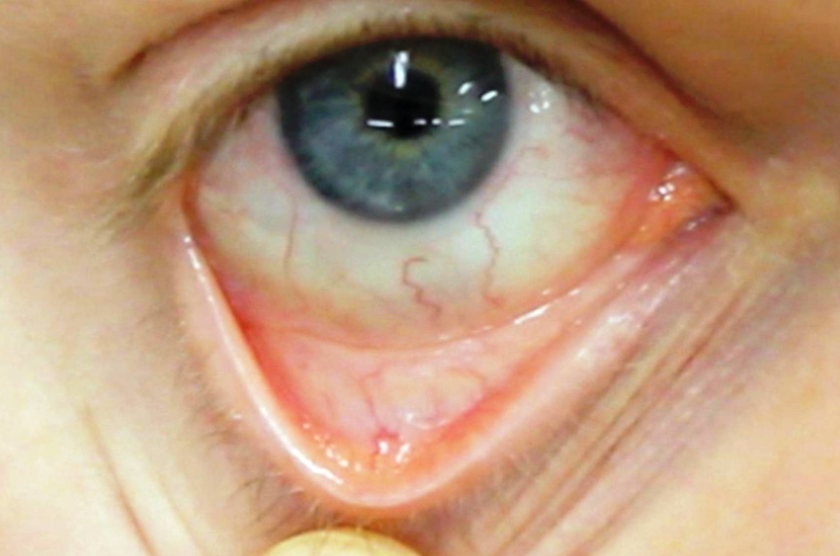
On examination, the signs include pallor of skin, conjunctiva and nail beds. Specific signs may be seen such as koilonychia (in iron deficiency), jaundice (in hemolytic anemia), bone deformities (in thalassemia major) or leg ulcers (in sickle-cell disease).
Lab Studies
- Complete blood count
- Examination of a well stained peripheral blood smear by a pathologist using a microscope
- Reticulocyte count
- ESR, ferritin, serum iron, transferrin, RBC folate level, serum vitamin B12, hemoglobin electrophoresis, renal function tests
- Bone marrow examination is an invasive procedure that allows direct examination of the red cell precursors. It is done in those cases where some specific pathology is under consideration.
Treatment of Anemia
Treatment depends on cause and severity. Identifying and treating the underlying cause remains the mainstay of the treatment.
An Overview of Treatment of Anemia
Oral therapy
Oral iron supplementation with ferrous sulfate, ferrous fumarate, or ferrous gluconate or vitamin supplements given orally (folic acid or vitamin).
Parenteral therapy
In cases where oral treatment has been ineffective, due to slow or impaired absorption, injectable iron or vitamin B12 can be used.
Blood transfusions
Blood transfusions are considered for patients having severe anemia.
Specific Treatment of Different Anemias
Anemia treatment depends on the cause.
Deficiency anemia
Includes anemia due to iron deficiency or vitamin deficiency
- Diet rich in iron
- Iron supplements – oral and/or parenteral
- Treatment of underlying cause. This may include medical or surgical treatment
Read more about 50 Iron Rich Foods You Can Add To Your Diet
Anemia of chronic disease
Apart from supportive treatment, the underlying cause should be treated. Supportive treatment includes diet changes, supplements, and blood transfusions if required.
Aplastic anemia
- Recurrent blood transfusion
- Bone marrow transplant
Anemias Associated with Bone Marrow Disease
- Medication
- Blood transfusion
- Chemotherapy
- Bone marrow transplantation.
Hemolytic anemias
- Stop medication causing hemolysis
- Treatment of infection (if infection is the cause)
- Blood transfusion
- Plasmapheresis
- Removal of spleen
Sickle cell anemia
- Administration of oxygen
- Analgesics
- Oral and intravenous fluids
- Blood transfusions
- Folic acid supplements
- Antibiotics
- Medication like hydroxyurea
Thalassemia
- Repeated blood transfusions
- Folic acid supplements
- Removal of the spleen
- Bone marrow transplant

